Duodenal Carcinoid
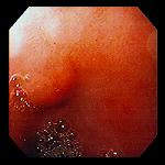
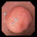
Left: 73 year-old woman with occult gastrointestinal bleeding was found to have this small, smooth mucosal nodule in the
duodenal bulb. Biopsies with special stains indicated this to be a carcinoid tumor; further workup was unremarkable, suggesting
this to be an isolated lesion.
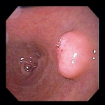
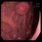
Right: Smooth sessile polypoid lesion in the duodenal bulb of a 71 year-old woman which proved to be carcinoid histologically;
there were no malignant features.
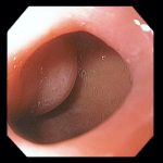
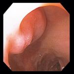
Incidental finding in a 76 year-old woman undergoing endoscopy for evaluation of abdominal pain. Smooth duodenal tumor visible through the pylorus on left. Closer view into the duodenal bulb on right.
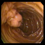
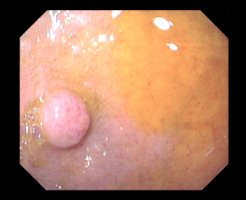
Left: 65 year old man undergoing endoscopy for dysphagia was found to have a 1 cm sessile polyp in the duodenal bulb.
Histology revealed a low-grade neuroendocrine (carcinoid) tumor with no high-grade features.
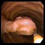
Right: 8-10 mm smooth submucosal lesion in the duodenal bulb of a 47 year old woman with abdominal pain. Histology revealed
a well-differentiated carcinoid tumor.
there were no malignant features.
53 year old woman with loss of appetite and weight loss. Endoscopy revealed an unusually prominent mucosal fold in the descending
duodenum, in the region of the ampulla; the ampulla itself was not visible. The fold was freely movable using a forceps, as seen in
the photo at right. Biopsies indicated the lesion to be, or the fold to contain, a well- differentiated carcinoid tumor.
70 year old woman with GERD, epigastric pain relieved by food ingestion, and a history of peptic ulcer with Helicobacter pylori positivity 3 years earlier. Endoscopic findings included this 4 mm smooth submucosal nodule in the anterior duodenal bulb. Biopsy revealed chronic duodenitis with a 2 mm focus of carcinoid at the base of the specimen. One month later she underwent endoscopic ultrasound and endoscopic mucosal resection of an 8 mm mass; histology showed a well-differentialed low-grade neuroendocrine tumor (carcinoid) which appeared to be superficial and completely excised. 10 months after the initial diagnosis, repeat endoscopic ultrasound and biopsies showed no evidence of residual disease.
1.5 years after the initial diagnosis she presented with epigastric pain radiating to the left upper abdominal quadrant, and back pain. She had no diarrhea, flushing or palpitations. CT scan revealed a left periaortic mass and mesenteric lymphadenopathy suggestive of metastatic disease. Octreotide scan revealed activity within the enlarged lymph nodes. There was no enhancement in the duodenum. She was placed on Sandostatin LAR. As of one year later (2.5 years after the initial diagnosis) her lesions were stable and symptoms were controlled on medication.
Endoscopic images Copyright © Atlanta South Gastroenterology, P.C. All rights reserved.
Logo is Registered Trademark ® of Atlanta South Gastroenterology, P.C.
This site is presented for educational and general informational purposes only. It does not purport to offer medical advice for any specific medical condition or individual patient. We regret that we cannot provide individualized medical advice online, either via this web site or via email. Please refer to our "Notable Web Sites" section, which offers links to several excellent online sources of additional medical information.