Lymphangiectasia
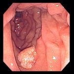
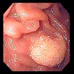
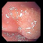
Left and Center: Focal lymphangiectasias in the postbulbar duodenum. Right: Close-up of image at center.
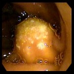
74 year old with abdominal pain and dyspepsia. Endoscopy revealed a 7-8 mm firm polypoid lesion in the second portion of the duodenum.
The surface villi appeared to be enlarged, possibly edematous. Biopsies revealed the lesion to be a lymphangioma, negative for adenoma
and malignancy.
Endoscopic images Copyright © Atlanta South Gastroenterology, P.C. All rights reserved.
Logo is Registered Trademark ® of Atlanta South Gastroenterology, P.C.
This site is presented for educational and general informational purposes only. It does not purport to offer medical advice for any specific medical condition or individual patient. We regret that we cannot provide individualized medical advice online, either via this web site or via email. Please refer to our "Notable Web Sites" section, which offers links to several excellent online sources of additional medical information.