Gastric Xanthelasma
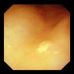
Left: Pale mucosal elevation found in the gastric antrum in a 76 year-old woman undergoing endoscopy for evaluation of bleeding. Biopsies revealed moderate chronic inflammation and increased numbers of foamy, lipid-laden histiocytes, consistent with a diagnosis of gastric xanthelasma (xanthoma). No Helicobacter pylori were seen.
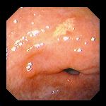
Right: Yellow mucosal plaque seen in the gastric antrum of a 53 year-old woman with acid-peptic symptoms that failed to respond to therapy. Biopsies revealed moderate to severe acute and chronic inflammation with numerous Helicobacter pylori, and collections of foamy macrophages underneath the surface epithelium, consistent with xanthelasma (xanthoma) of the stomach.
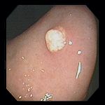
Left: Small polypoid xanthoma, with a pattern of fine pale granularity; an incidental finding at the time of percutaneous endoscopic gastrostomy placement in an 89 year-old woman with inability to swallow.
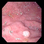
Right: Similar lesion in an 80 year old woman.
Endoscopic images Copyright © Atlanta South Gastroenterology, P.C. All rights reserved.
Logo is Registered Trademark ® of Atlanta South Gastroenterology, P.C.
This site is presented for educational and general informational purposes only. It does not purport to offer medical advice for any specific medical condition or individual patient. We regret that we cannot provide individualized medical advice online, either via this web site or via email. Please refer to our "Notable Web Sites" section, which offers links to several excellent online sources of additional medical information.