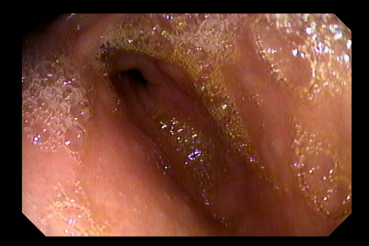
Intramucosal ("Early") Gastric Cancer
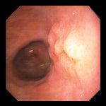
Left: 72 year-old man undergoing endoscopy for evaluation of abdominal pain. Endoscopy revealed a subtle, 2-3 cm focus of barely raised, nodular mucosa with normal color, on the posterior wall of the distal gastric body. The antrum and pylorus are visible in the background of this photo. Biopsies from the lesion revealed adenocarcinoma, and the patient was referred for surgical resection. The surgical specimen showed the lesion to be intra-mucosal, with invasion of the lamina propria but not into the muscularis mucosa.
Right: 67 year-old woman with unexplained weight loss as her only symptom. Endoscopy demonstrated mucosal irregularity with erosion and some thickening, but no frank ulceraton. Biopsy revealed adenocarcinoma. At surgery, the malignancy involved the mucosa and submucosa but did not invade the muscular layer. Lymph nodes were negative.
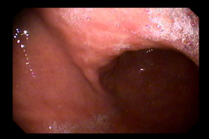
Left: 76 year-old woman who had undergone chemotherapy for a gastric MALT lymphoma, was found on post-treatment endoscopy to have a subtle raised lesion which ultimately proved to be a localized adenocarcinoma. There was no evidence of residual lymphoma.
Right: Same lesion as in center image, two weeks later at repeat endoscopy to obtain confirmatory biopsies. Residual signs of having been biopsied two weeks earlier can be seen.
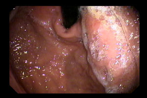
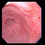
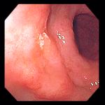
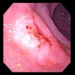
48 year old woman with significant weight loss and two weeks of upper abdominal pain.
Left: Endoscopy revealed poor distensibility of the gastric body, particularly the lesser curvature.
Center: Forward view of the lesser curvature showing an area of benign-appearing nodular mucosa.
Right: Same area of poor-distensibility and nodular mucosa as seen on retroflexed view.
Biopsies revealed adenocarcinoma, diffuse signet ring cell type, with a background of atrophic gastritis, intestinal metaplasia and Helicobacter pylori. CT scan with oral and intravenous contrast was read as normal, with no evidence of extension or metastasis. Endoscopic ultrasound suggested this to be early gastric cancer, T1M0, potentially curable with surgery.
Endoscopic images Copyright © Atlanta South Gastroenterology, P.C. All rights reserved.
Logo is Registered Trademark ® of Atlanta South Gastroenterology, P.C.
This site is presented for educational and general informational purposes only. It does not purport to offer medical advice for any specific medical condition or individual patient. We regret that we cannot provide individualized medical advice online, either via this web site or via email. Please refer to our "Notable Web Sites" section, which offers links to several excellent online sources of additional medical information.